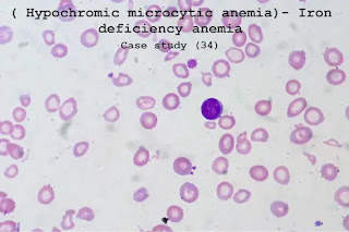
Case study (34) –( Hypochromic microcytic anemia)- Iron deficiency anemia
A 72-year-old woman with a medical history of 4 to 6 weeks gradually increased fatigue and shortness of breath.
Laboratory Investigations:
Hemoglobin (Hb) 86 g/L
Mean corpuscular volume (MCV) 65 fL
Mean corpuscular hemoglobin (MCH) 26.4 pg
Mean corpuscular hemoglobin concentration (MCHC) 29 g/dL
Red blood cells (RBC) 3.8 X 10¹²/L
White blood cells (WBC) 6.7 X 10⁹/L (differential normal)
Platelets 456 X 109/L
Questions:
Q1. What abnormalities are shown in the blood film?
Q2. What is the diagnosis?
Q3. What further investigations should be performed?
Answers:
A1. The blood count shows anemia with microcytic, hypochromic red cell indices.
A2. The platelet count is raised, in keeping with iron deficiency anemia.
The blood film shows hypochromic, and the number of microcytic cells, target cells, and high numbers of the platelet.
The differential diagnosis of hypochromasia and microcytosis is thalassemia.
The carrier states for both alpha-thalassemia and beta-thalassemia are rarely associated with anemia.
Other causes of target cells include liver disease, post-splenectomy, and other hemoglobinopathies, for example, hemoglobin C.
A3. Investigations should aim at confirming iron deficiency and defining a cause.
Reduce serum iron, increase iron-binding capacity, and reduce serum ferritin (this is an accurate measure of iron reserves in the body).
The most common cause of this picture is bleeding. In postmenopausal women or men, the cause of bleeding must be determined.
In this patient, a barium enema was performed.
This appeared a carcinoma of the colon at the splenic flexure.


Comments
Post a Comment